One in 10,000 newborns is born with malformations of the bladder, intestines or genitals. These symptoms are part of the so-called bladder exstrophy epispadias complex, abbreviated BEEC. Since the disorder tends to run in families, it is assumed to have a genetic cause. However, up to now there has been disagreement as to exactly which genetic material is affected or whether there are even several genes involved.
The recently published study sheds light on this issue. Four years ago, researchers led by Prof. Dr. Heiko Reutter from the Institute of Human Genetics at the University of Bonn discovered a gene that is abnormal in sick children. The gene bears the cryptic abbreviation SLC20A1. "We have now taken a closer look at its function," explains Magdalena Rieke, who is completing her doctorate under Prof. Reutter.
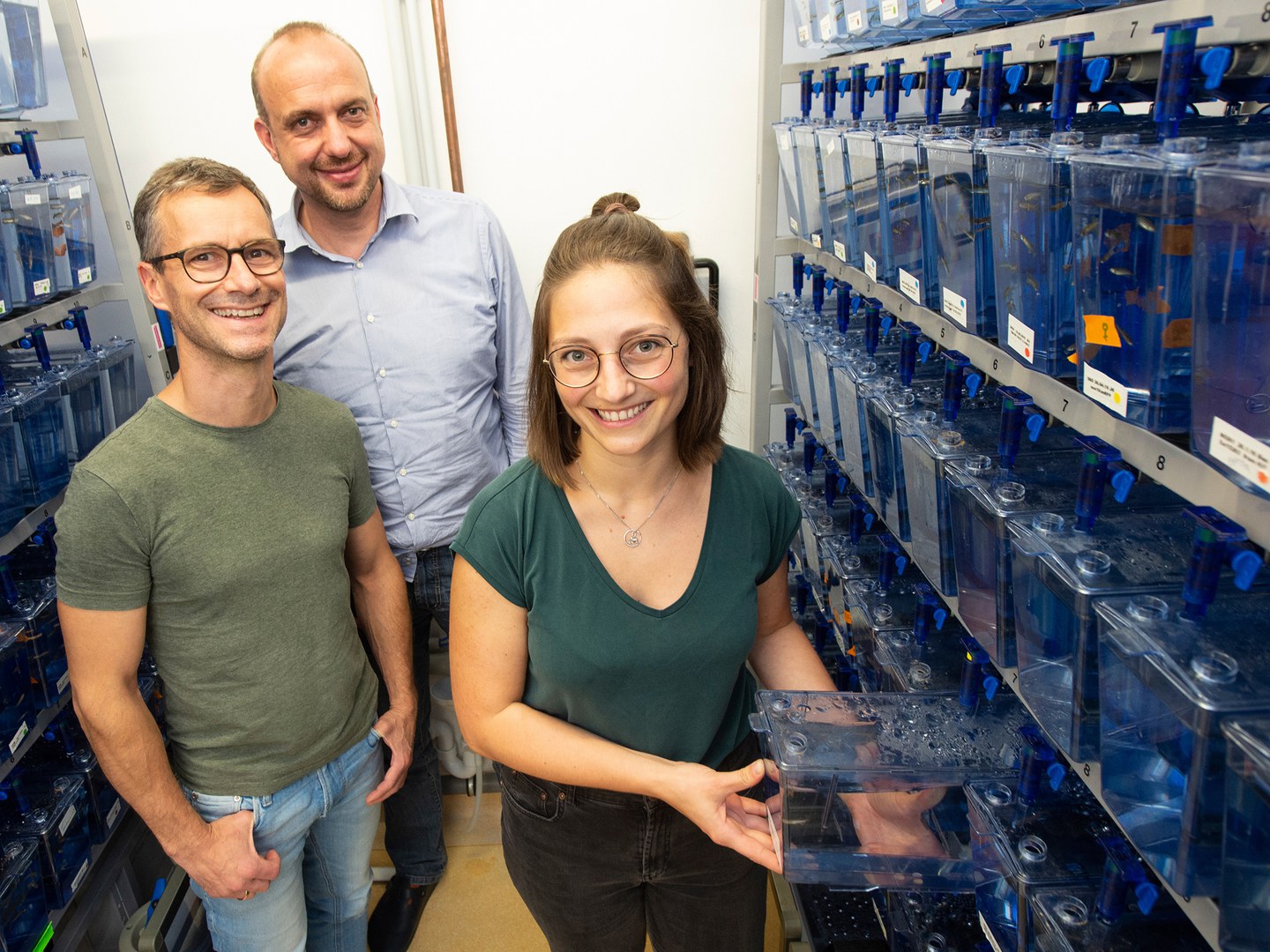
The researcher also benefited from the expertise of a university working group that only marginally deals with congenital malformations: Prof. Dr. Benjamin Odermatt researches the cause of neurological diseases at the Section of Neuroanatomy. The zebrafish serves as a model organism. Not only because it can be easily kept in an appropriate habitat and reproduces quickly: Many of its genes are also found in a very similar form in humans.
Zebrafish as genetic model
This also includes SLC20A1. "We used an active substance in the animals to prevent the gene from being translated into proteins," explains Rieke. "As a result, the growing larvae showed disrupted development of their excretory organs. This indicates that SLC20A1 really does appear to play a central role in the correct formation of these organs, and has done so for many millions of years." The researchers were furthermore able to show that the gene is also active in human embryos, particularly in structures involved in the formation of the excretory organs and genitalia.
In human patients, the researchers found three different mutations of SLC20A1. These anomalies often occur spontaneously. Therefore, even children whose parents are completely healthy may be affected. Rieke and her colleagues were able to demonstrate the effect of one of these mutations in human cell cultures: It interferes with the controlled degradation of cells, the "programmed cell death", a very important step in tissue remodeling.
During embryonic development, not only are masses of new cells produced, but some are also deliberately destroyed. This is for instance how the opening of the intestine to the outside, the anus, is created. Researchers refer to the process of programmed cell death as apoptosis. "This association might explain why mutations in SLC20A1 can cause such severe developmental disorders," speculates Rieke.
Impaired protein folding
SLC20A1 contains the building instructions of a protein that is located in the cell membrane, the fat-like envelope that surrounds the cells. This protein resembles a long worm that has arranged its body in numerous tight loops that repeatedly run from the outside of the membrane to the inside and back. Computer models suggest that at least one of the mutations discovered prevents correct folding. This is thought to severely disrupt protein function, and thus also the activation of apoptosis.
It is not yet possible to derive immediate insights for the treatment of BEEC directly from the results. "However, it is essential that we gain a better understanding of the disease mechanism for any possible prevention or therapy," stresses Rieke, who herself works as an assistant doctor in the field of pediatric and adolescent medicine. In addition to various working groups from Bonn and Germany, research institutions from Sweden, Great Britain, Italy, India and the Netherlands were also involved in the study. It is thus also an example of successful international cooperation.
Publication: Johanna Magdalena Rieke and others: SLC20A1 is involved in urinary tract and urorectal development. Frontiers in Cell and Developmental Biology, DOI: 10.3389/fcell.2020.00567
Contact:
Magdalena Rieke and Prof. Dr. Heiko Reutter
Institut für Humangenetik, Universitätsklinikum Bonn
Tel. +49(0)-228-6885419
E-mail: magdalena.rieke@ukbonn.de,reutter@uni-bonn.de
Prof. Dr. Benjamin Odermatt
Institut für Anatomie, Universität Bonn
Tel. +49(0)-228-739021
E-mail: b.odermatt@uni-bonn.de


