Disease-causing bacteria of the genus Salmonella or Yersinia can use tiny injection apparatuses to inject harmful proteins into host cells, much to the discomfort of the infected person. However, it is not only with a view to controlling disease that researchers are investigating the injection mechanism of these so-called type III secretion systems, also known as "injectisomes". If the structure and function of the injectisome were fully understood, researchers would be able to hijack it to deliver specific drugs into cells, such as cancer cells. In fact, the structure of the injectisome has already been elucidated. However, it remained unclear how the bacteria load their syringes so that the right proteins are injected at the right time.
A team of scientists led by Dr. Andreas Diepold from the Max Planck Institute for Terrestrial Microbiology in Marburg and Prof. Dr. Ulrike Endesfelder from the University of Bonn has now been able to answer this question: mobile components of the injectisome comb through the bacterial cell in search of the proteins to be injected, so-called effectors. When they encounter an effector, they transport it like a shuttle bus to the gate of the injection needle. "How proteins of the sorting platform in the cytosol bind to effectors and deliver the cargo to the export gate of the membrane-bound injectisome is comparable to the processes at a freight terminal", explains Dr. Stephan Wimmi, one of the first authors of the study as a postdoctoral researcher in Andreas Diepold's laboratory. "We think that this shuttle mechanism helps to make the injection efficient and specific at the same time - after all, the bacteria have to inject the right proteins quickly to avoid being recognized and eliminated by the immune system, for example."
To gain this insight into the important loading mechanism of the injectisome, the researchers had to apply new techniques. "Conventional methods, which are normally used to detect that proteins bind to each other, did not work to answer this question - possibly because the effectors are only bound for a short time and then immediately injected," explains Andreas Diepold, research group leader at the Max Planck Institute and co-leader of the study. "That's why we had to analyse this binding in situ in the living bacteria.”
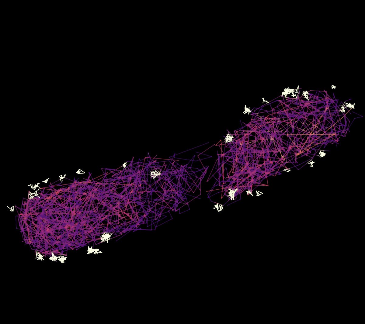
“To measure these transient interactions we made use of two novel approaches that work in living cells, proximity labeling and single-particle tracking, ” adds Ulrike Endesfelder, a member of the Transdisciplinary Research Areas “Modelling” and “Life and Health” at the University of Bonn. Her group worked on the study in three different locations – the MPI in Marburg, Carnogie Mellon University in Pittsburgh, USA, and at the University in Bonn. “Proximity labeling—a technology in which a protein marks its immediate neighbors, as if with a brush—revealed that the effectors in the bacterium bind to the mobile injectisome components.” Her team then studied this binding via the high-resolution microscopy method of single particle tracking. “We are able to employ this method to track individual proteins,” explains University of Bonn doctoral student Alexander Balinovic, who is a lead author of the study. “We mark the components we intend to track with photoactivatable fluorescent proteins, which then enable us to localize the individual particles in microscopy imaging.” These methods, which the team refers to as "in situ biochemistry", i.e. biochemical investigations on site, made the breakthrough possible, which has now been published in the journal "Nature Microbiology".


