Chronically stressed people often show abnormalities in their motor skills, such as poorer fine motor control. However, how these symptoms occur has hardly been studied so far. "We investigated this question in our study," explains Prof. Dr. Valentin Stein from the Institute of Physiology II at the University of Bonn.
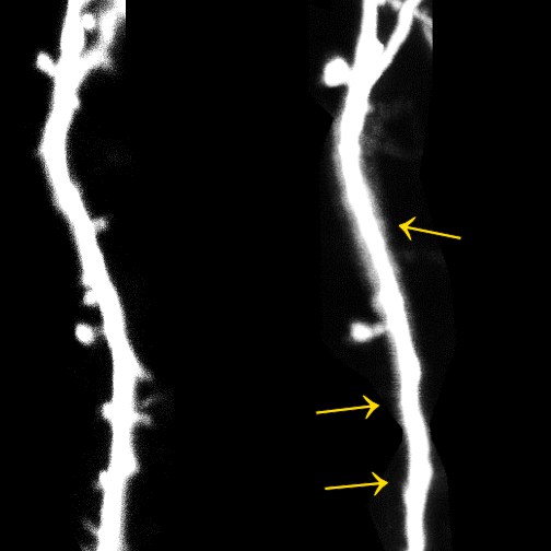
The researchers used mice as experimental animals, some of which they exposed to a stressful situation for a few days. Meanwhile, they used a special microscopy method to take pictures of the rodents' brains. They focused on parts of the cerebral cortex responsible for motor control and learning new movements.
"With our method, it is possible to observe one and the same neuron at different points in time," says Dr. Anne-Kathrin Gellner, a physician at the Department of Psychiatry and Psychotherapy at Bonn University Hospital. "We can therefore see whether and how it changes as a result of stress."
Stressed mice lose synapses
In fact, the researchers came across a conspicuous feature: after the stressful situation, the neurons studied lost some of their synapses - these are the contacts to other nerve cells. During learning processes, new synapses are usually formed or existing ones are strengthened. Instead, the stressed rodents lost up to 15 percent of their contacts.
At the same time, the animals developed motor learning deficits. For example, they had to try to grasp a food pellet with one paw and transport it into their mouths. In the wild, mice use both paws to do this, so they had to relearn this skill. The non-stressed control group achieved a success rate of 30 percent after five days. The stressed rodents, however, only managed to take the food in every tenth attempt.
Mice vary in their sensitivity to stress. Some of them hardly develop any abnormalities after a few days of stress - they are considered resilient. Surprisingly, these robust animals had similar difficulties as their more sensitive peers in learning to grasp with one hand. "It is therefore possible that motor tests are very suitable for detecting stress-related disorders such as depression before other symptoms become apparent," hopes Prof. Valentin Stein.
Even resilient animals are not immune
Even in resilient animals, moreover, the number of synapses decreased after the stress event. Unlike their stress-sensitive peers, however, the affected neurons recovered: after one and a half weeks, the number of synapses was again similar to that before the stress event and comparable to that in non-stressed control animals. "Nevertheless, it may well be that psychological stress also leaves permanent traces on them if it is too long or too frequent," worries Stein, who is also a member of the Transdisciplinary Research Area (TRA) "Life and Health."
The researchers also have clues as to what triggers the loss of synapses: Certain immune cells, the microglia, were activated in the rodents' brains. They belong to the so-called phagocytes and can, for example, digest pathogens or defective cells. It is possible that they are "switched on" by stress and then attack the contact sites.
The research group also examined the fluid that washes around the brain and spinal cord. They found certain proteins that can normally be detected there in neurodegenerative diseases such as Parkinson's or Alzheimer's. "We therefore believe that stress-related psychiatric diseases such as depression are also associated with the degradation of neurons," says Dr. Gellner. "Accordingly, long-term stress - to which children are increasingly exposed - can potentially cause serious damage to the brain."
Participating institutions and funding:
The Department of Psychiatry and Psychotherapy of the University Hospital Bonn, the Institute of Nutritional Research of the University of Potsdam, the Institute of Biochemistry and Molecular Biology, the Institute of Molecular Psychiatry, and the Institute of Physiology II (all University of Bonn) were involved in the study. Dr. Anne-Kathrin Gellner was supported by the BONFOR program of the Faculty of Medicine.
Publication: Anne-Kathrin Gellner, Aileen Sitter, Michal Rackiewicz, Marc Sylvester, Alexandra Philipsen, Andreas Zimmer and Valentin Stein: Stress vulnerability shapes disruption of motor cortical neuroplasticity. Translational Psychiatry, DOI: 10.1038/s41398-022-01855-8.
Contact:
Prof. Dr. Valentin Stein
Institute of Physiology II
University of Bonn
Phone +49-(0)228-7360157
E-mail: vstein@uni-bonn.de


